
Aslanger Pattern
Item from last night and a reminder from our EKG STEMI-equivalent lecture.
The Aslanger Pattern
Why it matters:
The Aslanger pattern shows up in roughly 6% of NSTEMI patients and is associated with larger infarct size and higher mortality. The danger is not subtle ECG findings. It’s that this is an OMI masquerading as a non-STEMI, leading to delayed cath.
What’s actually happening:
This is an occlusion MI pattern, most often due to critical RCA or LCx stenosis in patients with multivessel coronary disease. Because there is competing ischemia across multiple territories, the ECG never develops classic contiguous ST-elevation and therefore does not meet STEMI criteria.
Why we miss it:
The ECG looks “NSTEMI-ish,” so cath is often delayed. But the myocardium is still acutely threatened.
The ECG pattern to recognize:
Isolated ST elevation in lead III
ST depression in at least one of V4–V6, with a positive or terminally positive T-wave
ST segment in V1 greater than V2
ECG Basics, no cardiology fellowship required:
Lead III and V1 are anatomically contiguous. Lead III and V1 are looking at nearby parts of the heart in the inferior–posterior region.
With inferior injury and lateral ischemia occurring simultaneously (due to multivessel disease), you get isolated elevation in III and ST V1 > V2, with reciprocal depression laterally.
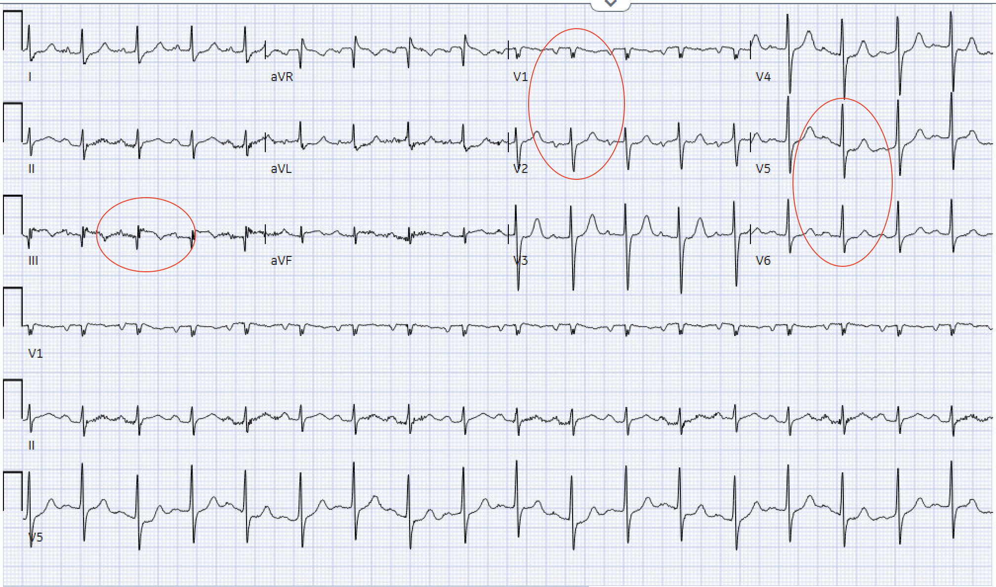
Clear Example of OMI with Aslanger Pattern from LITFL:
(1) STE in III but not in any other inferior lead
(2) ST depression in any of leads V4 to 6 with a positive (at least terminally positive) T-wave
(3) ST in lead V1 higher than ST in V2
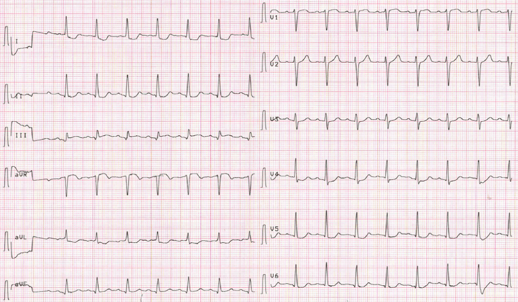
EKG from yesterday evening.
(1) STE in III but not in any other inferior lead
(2) ST depression in any of leads V4 to 6 with a positive (at least terminally positive) T-wave
(3) ST in lead V1 higher than ST in V2
The ECG from yesterday evening looks to fit this pattern (but might be an over-read on my part). The patient underwent PCI and was found to have a 95% distal RCA culprit lesion, now stented, with multivessel disease. This finding is consistent with patients noted to have Aslanger pattern in the original study.
Bottom line
- If you see isolated STE in III with lateral ST depression and V1>V2, in a patient that screams MI, then this is Aslanger. This is an inferior OMI until proven otherwise.
Read More About This:
References:
Aslanger E, Yıldırımtürk Ö, Şimşek B, Sungur A, Türer Cabbar A, Bozbeyoğlu E, Karabay CY, Smith SW, Değertekin M. A new electrocardiographic pattern indicating inferior myocardial infarction. J Electrocardiol. 2020 Jul-Aug;61:41-46. [Pubmed]
Cheers,
Dillon